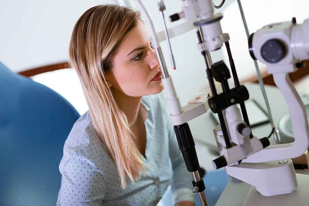
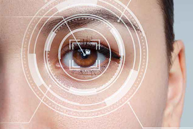
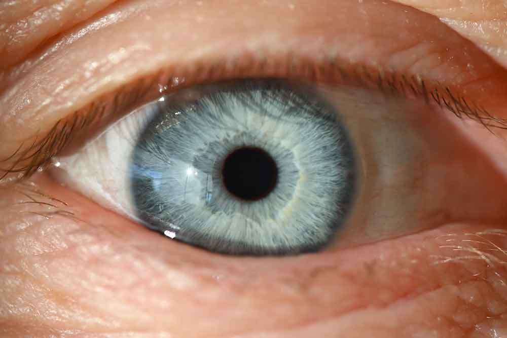
Visual Field Test
When you focus your attention on something, a certain portion of your visual field becomes visible to you. For instance, when you look at a car, your eyes are directed toward the car, but you may also notice things that are in the immediate vicinity of the car, such as flowers growing in the ground or raindrops falling on the windshield. Your capacity to see what is in this immediate region will be evaluated using a visual field test. As a result of the potential for certain eye conditions as well as neurological disorders to have a negative impact on a person's visual field, this test is an essential component of a comprehensive eye exam.
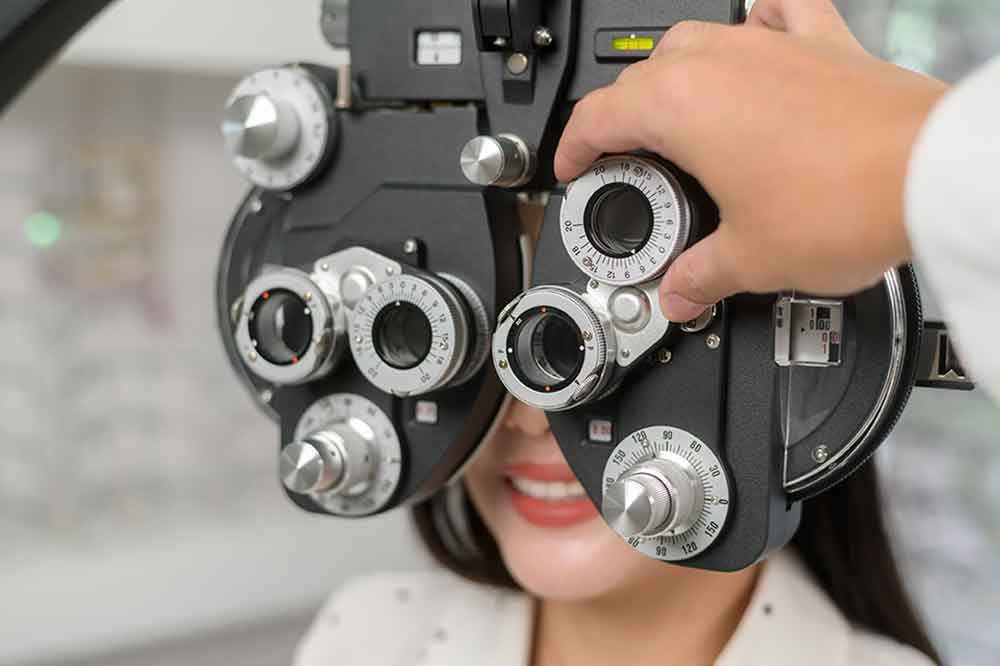
Examination of Refraction
A standard eye and vision exam typically includes a refraction exam as one of its component tests. The way you see the world around you is called refraction. Vision that is blurry is the result of an imperfection in the refraction of your eye, which can occur at any age. Both nearsightedness and farsightedness are the most common types of vision problems caused by refractive errors. This straightforward examination may be carried out by the physician by shining a light into your eyes in order to observe how the light bends through it; alternatively, a computerized test may also be utilized.
Binocular Vision Assessment
Binocular vision is the ability of both of your eyes to focus on the same image or object so that your brain is able to "translate" what you are seeing from the image into an understanding of what you are seeing. This is analogous to how binoculars allow you to use both eyes simultaneously when looking at something far away. Binocular vision refers, at its most fundamental level, to the way in which the two eyes collaborate to form a single image.
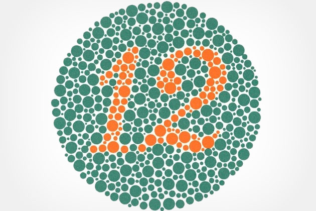
Assessment of Color
The ability to distinguish one hue from another is put to the test during a color assessment. Ishihara color plates are typically used for this purpose. These plates are a series of round circles with colored dots inside that form a number. The patient examines the image, which may be displayed on paper or on the screen of a computer, and determines whether or not they can make out the number. This is an essential test because having trouble telling the difference between red and green is one of the most common symptoms of color blindness.
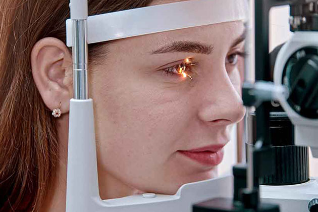
Eye Pressure Test
An eye pressure test, also known as a tonometry test, measures the amount of intraocular pressure (IOP) that is present in your eye at the time of the examination. The evaluation of eye pressure is an indispensable component of any comprehensive eye exam. Glaucoma is one of the conditions that can be indicated by abnormally high eye pressure. This examination begins with eye drops that numb the eye, followed by the application of a small device that the doctor uses to lightly touch the eye while evaluating the patient's intraocular pressure.
Evaluation of Peripheral Vision
The ability to see what is to the side without moving your head is referred to as having peripheral vision. Even though your eyes may be directed toward a particular image or object, you are still able to see things in the surrounding area. The doctor will perform this evaluation by giving you instructions to concentrate on something that is right in front of you, like a pen, for example. You will then be asked what you can see to the side or a little bit further away from that object. This test can determine whether or not there is any loss of peripheral vision, which can be an indication of a number of different eye diseases, such as glaucoma, diabetic retinopathy, or retinal pigmentosa.
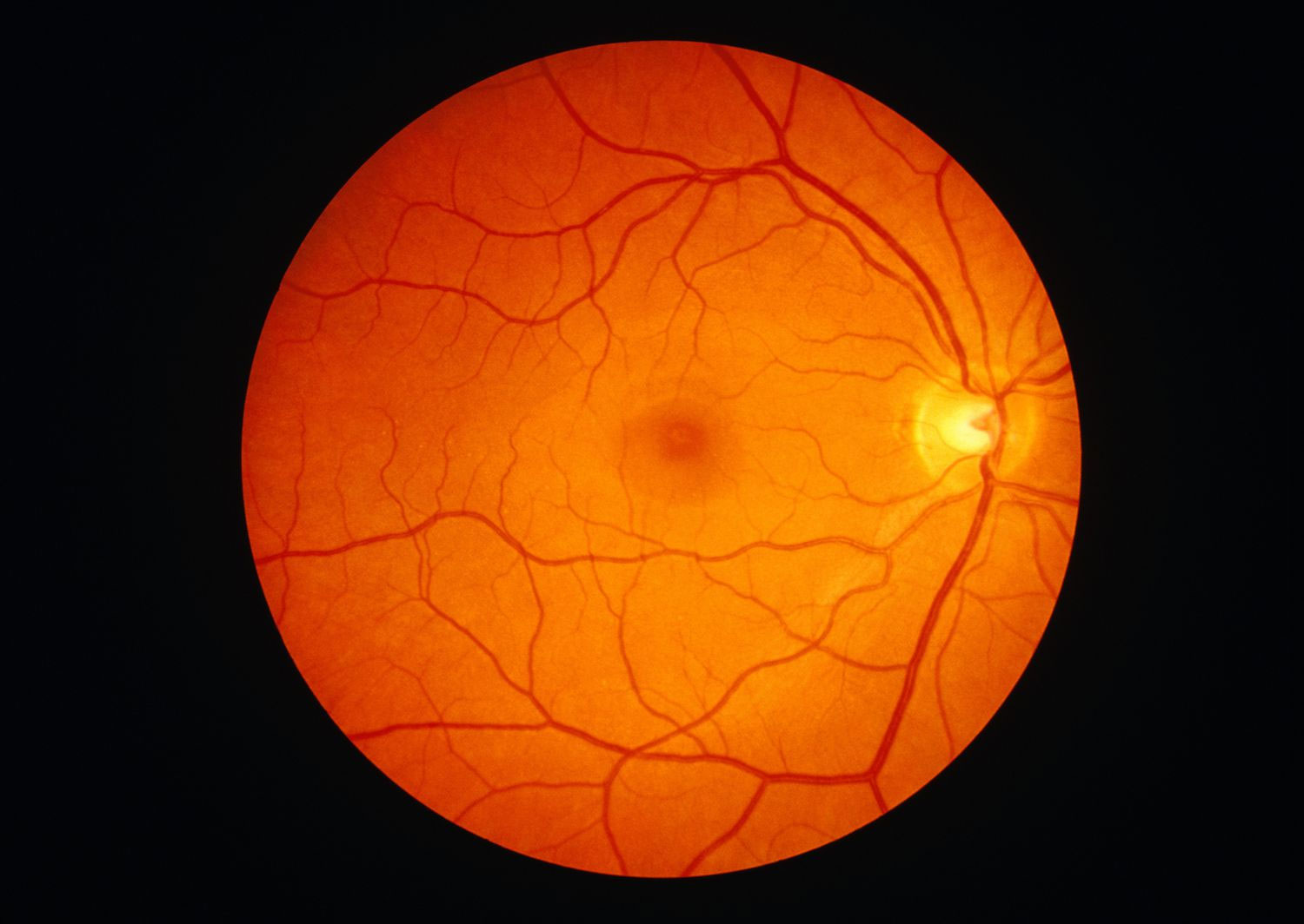
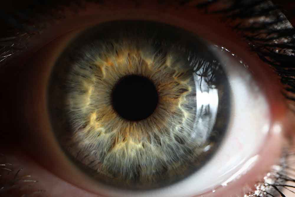
Digital Retinal Imaging
The technology of digital retinal imaging gives your doctor the ability to examine the condition of your retina. The photograph was taken with a specialized kind of digital camera. The image of the retina, which is found in the interior of the eye's posterior chamber, is captured by the camera and saved in an electronic format. This is essential to your vision because the retina helps focus light that enters your eye and sends images to the brain, which ultimately allows you to comprehend what you are seeing.
Mapping of the Cornea
Measurement of the cornea is an important part of the process known as corneal mapping. In order to obtain precise information regarding the dimensions and contours of your cornea, we will use either a computerized system or a keratometer. This is done to ensure that the curvature and size are correct, which enables light to enter your eye, allowing you to focus on images and see clearly. This is done to ensure that the cornea has the correct curvature and size.
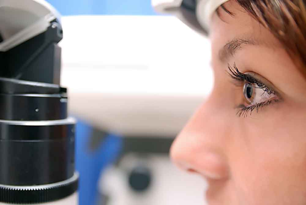
OCT Scan
An OCT scan, also known as an optical coherence tomography scan, is a diagnostic procedure that, like a traditional CT scan, examines the layers of your retina and optic nerve to look for signs of eye diseases. This examination requires the use of a laser that emits light in order to produce high-resolution, color images of the retina for the examining physician. The examination is non-invasive, does not cause any pain, and does not use any radiation.
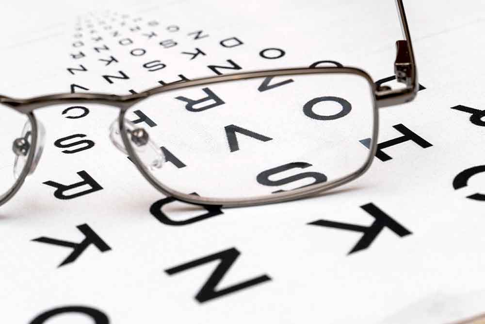
The Test of Visual Acuity
The ability to see images that are clear and distinct from a variety of distances is known as visual acuity. The physician will have you look at an eye chart while being exposed to a variety of different intensities of light in order to evaluate your proficiency in this area. Your level of visual acuity can be determined by the smallest letters or numbers that you are able to see clearly.